


Die Hornhauttransplantation
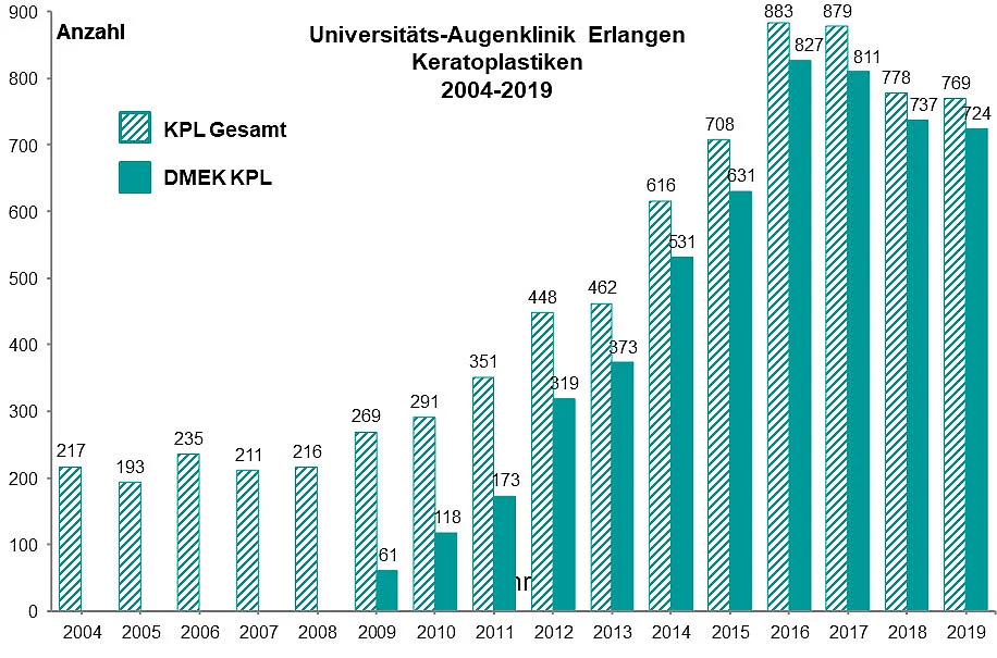
Die Hornhauttransplantation ist die am häufigsten durchgeführte und erfolgreichste Transplantation in der gesamten Medizin. Die Erlanger Universitäts-Augenklinik hat im vergangenen Jahr 778 Hornhauttransplantationen (Keratoplastiken) durchgeführt und ist damit führend in Deutschland und in Europa. Damit nehmen wir auf diesem Gebiet in Europa und weltweit mit Abstand die Spitzenposition ein. Insbesondere kommen an unserer Klinik die modernen Verfahren der schichtweisen Hornhautübertragung (DMEK, DSAEK und DALK) zum Einsatz. Bislang haben wir über 6000 Hornhauttransplantationen nach diesem modernen Verfahren durchgeführt.
Die Erlanger Augenchirurgen um Prof. Dr. Friedrich E. Kruse entwickeln die Technik der Hornhauttransplantation seit fast 20 Jahren weiter und haben vor 15 Jahren die neuen lamellären Techniken, insbesondere die DMEK und DSAEK in Deutschland eingeführt. Die Hornhauttransplantation als DMEK wird an der Universitäts-Augenklinik Erlangen mittlerweile bei Patienten mit Fuchs’scher Hornhautdystrophie und anderen Erkrankungen des Hornhautendothels als Standardverfahren angewandt.
In den letzten 15 Jahren fand eine sprunghafte Entwicklung im Bereich der Hornhauttransplantation statt, die es heutzutage ermöglicht, einzelne Schichten der Hornhaut zu ersetzen. Ein Paradebeispiel dieser Methode ist die DMEK, bei der das Endothel ersetzt wird. Der Oberbegriff dieser Techniken ist die lamelläre Keratoplastik. Bei der lamellären Hornhauttransplantation unterscheidet man grundsätzlich zwischen Operationen, bei denen die Oberfläche und der mittlere Teil der Hornhaut (Epithel und Stroma) ersetzt werden (vordere lamelläre Keratoplastik [DALK]) und Operationen, bei denen nur die innerste Schicht, also das Endothel ersetzt wird (hintere lamelläre Keratoplastik [DMEK und DSAEK]).
Was ist die Hornhaut?
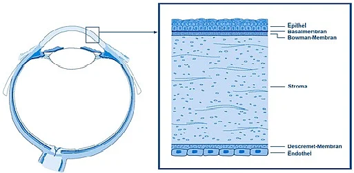
Die Hornhaut ist das klare Fenster des Auges (Abbildung1). Sie schließt den runden Augapfel nach vorne ab, ähnlich wie das Glas einer Uhr (Abbildung 2). Nur wenn die Hornhaut durchsichtig und gewölbt ist, kann man scharf sehen.
Wie ist die Hornhaut aufgebaut?
Die Hornhaut ist ein klares durchsichtiges Scheibchen mit einem Durchmesser von 11 mm. Sie ist nur ½ mm dick. Wie bei einer Fensterscheibe unterscheidet man auch bei der Hornhaut eine Außenschicht, einen mittleren Teil und eine Innenschicht. In der Fachsprache bezeichnet man die Außenschicht als das Epithel, die mittlere Schicht als das Stroma und die innere Schicht als das Endothel. Das Besondere an der Endothelzellschicht ist, dass sie kleine Pumpen besitzt, die Wasser aus der Hornhaut herauspumpen. Nur, wenn diese Pumpen funktionieren, kann man klar sehen. Die Endothelzellen sitzen auf einem papierdünnen Häutchen, der so genannten Descemt’schen Membran.
Was gibt es für Hornhauterkrankungen?
Jede Schicht der Hornhaut kann eine eigene Erkrankung entwickeln. Die meisten kann man mit Medikamenten behandeln. Entwickeln sich durch Infektionen oder Verletzungen jedoch Hornhauttrübungen und Narben, müssen diese operiert werden. Zu den Erkrankungen der mittleren Hornhautschicht gehören Veränderungen, die die Form der Hornhaut verändern, wie der Keratokonus oder Verdünnungen am Rand der Hornhaut. Die Erkrankungen der Innenschicht (Endothel) führen ebenfalls zur Eintrübung, dazu gehört die Fuchs’sche Hornhaut-Endothel-Dystrophie und die pseudophake bullöse Keratopathie nach Grauer Star Operation.
Kann man die Hornhaut operieren - oder - was ist eine Hornhauttransplantation?
Der Austausch einer krankhaft veränderten Hornhaut im Rahmen einer Operation bezeichnet man als Hornhauttransplantation oder Keratoplastik. Die Transplantation der Hornhaut am Auge ist die häufigste Gewebetransplantation beim Menschen. Pro Jahr werden etwa 5.000 Spenderhornhäute verpflanzt. Die Universitäts-Augenklinik Erlangen ist das größte Transplantationszentrum für Hornhauttransplantationen in Deutschland. Hier wurden deutschlandweit auch die erste DSAEK- und insbesondere die erste DMEK durchgeführt. Die Universitäts-Augenklinik Erlangen nimmt weltweit einen Spitzenplatz in der Erforschung der neuen Transplantationstechniken ein. An keiner anderen Universitäts-Augenklinik in Deutschland sind in den letzten Jahren so viele Artikel zu den neuen Techniken DALK, DSAEK, DMEK publiziert wie an der Erlanger Universitätsaugenklinik. Eine Auswahl unserer Arbeiten finden Sie hier:
Bei Bedarf senden wir Ihnen gerne einen Sonderdruck zu.
Auszug aus der Publikationsliste zum Thema Hornhauttransplantation (DMEK, DSAEK, DALK und perforierende Keratoplastik)
Publikationen der Erlanger Arbeitsgruppe
Schlötzer-Schrehardt U, Zenkel M, Strunz M, Gießl A, Schondorf H, da Silva H, Schmidt GA, Greiner MA, Okumura N, Koizumi N, Kinoshita S, Tourtas T, Kruse FE. Potential Functional Restoration of Corneal Endothelial Cells in Fuchs Endothelial Corneal Dystrophy by ROCK Inhibitor (Ripasudil). Am J Ophthalmol. 2020 Dec 11; 224:185-199. Epub ahead of print.
Augustin VA, Weller JM, Kruse FE, Tourtas T. Refractive Outcomes After Descemet Membrane Endothelial Keratoplasty + Cataract/Intraocular Lens Triple Procedure: A Fellow Eye Comparison. Cornea. 2020 Nov 9. Epub ahead of print.
Augustin VA, Weller JM, Kruse FE, Tourtas T. Influence of corneal guttae and nuclear cataract on contrast sensitivity. Br J Ophthalmol. 2020 Sep 9. Epub ahead of print.
Lau N, Hajjar Sesé A, Augustin VA, Kuit G, Wilkins MR, Tourtas T, Kruse FE, Højgaard-Olsen K, Manuel R, Armitage WJ, Larkin DF, Tuft SJ. Fungal infection after endothelial keratoplasty: association with hypothermic corneal storage. Br J Ophthalmol. 2019 Oct;103(10):1487-1490.
Menzel-Severing J, Walter P, Plum WJ, Kruse FE, Salla S. Assessment of Corneal Endothelium during Continued Organ Culture of Pre-Stripped Human Donor Tissue for DMEK Surgery. Curr Eye Res. 2018 Dec;43(12):1439-1444.
Augustin VA, Weller JM, Kruse FE, Tourtas T. Fungal Interface Keratitis After Descemet Membrane Endothelial Keratoplasty. Cornea. 2018 Nov;37(11):1366-1369.
Augustin VA, Weller JM, Kruse FE, Tourtas T. Can we predict the refractive outcome after triple Descemet membrane endothelial keratoplasty? Eur J Ophthalmol. 2019 Mar;29(2):165-170.
von Marchtaler PV, Weller JM, Kruse FE, Tourtas T. Air Versus Sulfur Hexafluoride Gas Tamponade in Descemet Membrane Endothelial Keratoplasty: A Fellow Eye Comparison. Cornea. 2018 Jan;37(1):15-19.
Akbaba Y, Weller JM, Rössler K, Armitage WJ, Schlötzer-Schrehardt U, Kruse FE, Tourtas T. "Bubble-in-the-Roll" Technique Using the Endoject DMEK Injector: Influence of the Air Bubble on Endothelial Cell Loss. Cornea. 2017 Dec;36(12):1576-1579.
Menzel-Severing J, Kruse FE, Tourtas T. Organ-cultured, prestripped donor tissue for DMEK surgery: clinical outcomes. Br J Ophthalmol. 2017 Aug;101(8):1124-1127.
Schmeckenbächer N, Frings A, Kruse FE, Tourtas T. Role of Initial Intraocular Pressure in Graft Adhesion After Descemet Membrane Endothelial Keratoplasty. Cornea. 2017 Jan;36(1):7-10.
Weller JM, Schlötzer-Schrehardt U, Kruse FE, Tourtas T. Splitting of the Recipient's Descemet Membrane in Descemet Membrane Endothelial Keratoplasty- Ultrastructure and Clinical Relevance. Am J Ophthalmol. 2016 Dec; 172:1-6.
Schlögl A, Tourtas T, Kruse FE, Weller JM. Long-term Clinical Outcome After Descemet Membrane Endothelial Keratoplasty. Am J Ophthalmol. 2016 Sep; 169:218-226.
Weller JM, Schlötzer-Schrehardt U, Tourtas T, Kruse FE. Influence of Ultrastructural Corneal Graft Abnormalities on the Outcome of Descemet Membrane Endothelial Keratoplasty. Am J Ophthalmol. 2016 Sep; 169:58-67.
Menzel-Severing J, Salla S, Plum WJ, Tourtas T, Fuchsluger T, Schlötzer- Schrehardt U, Kruse FE. Instrument to Enhance Visualization of Descemet Membrane During Graft Preparation for DMEK Surgery. Cornea. 2016 Jun;35(6):904-7.
Ćirković A, Beck C, Weller JM, Kruse FE, Tourtas T. Anterior Chamber Air Bubble to Achieve Graft Attachment After DMEK: Is Bigger Always Better? Cornea. 2016 Apr;35(4):482-5.
Weller JM, Tourtas T, Kruse FE. Feasibility and Outcome of Descemet Membrane Endothelial Keratoplasty in Complex Anterior Segment and Vitreous Disease. Cornea. 2015 Nov;34(11):1351-7.
Menzel-Severing J, Petsch C, Tourtas T, Polisetti N, Klenke J, Skerl K, Wüllner C, Donitzky C, Kruse FE, Kremers J, Hammer CM. Evaluation of a 345 nm Femtosecond Laser for Corneal Surgery with Respect to Intraocular Radiation Hazard. PLoS One. 2015 Sep 11;10(9):e0137638.
Tourtas T, Weller JM, Bachmann BO, Kruse FE. Larger Descemetorhexis to Improve Graft Adhesion in Descemet Membrane Endothelial Keratoplasty Does Not Cause Postoperative Peripheral Corneal Edema. Eye Contact Lens. 2015 Nov;41(6):344-8.24.
Kruse FE, Schrehardt US, Tourtas T. Optimizing outcomes with Descemet's membrane endothelial keratoplasty. Curr Opin Ophthalmol. 2014 Jul;25(4):325-34.
Price MO, Price FW Jr, Kruse FE, Bachmann BO, Tourtas T. Randomized comparison of topical prednisolone acetate 1% versus fluorometholone 0.1% in the first year after descemet membrane endothelial keratoplasty. Cornea. 2014 Sep;33(9):880-6.
Weller JM, Tourtas T, Kruse FE, Schlötzer-Schrehardt U, Fuchsluger T, Bachmann BO. Descemet membrane endothelial keratoplasty as treatment for graft failure after descemet stripping automated endothelial keratoplasty. Am J Ophthalmol. 2015 Jun;159(6):1050-1057.
Schlötzer-Schrehardt U, Bachmann BO, Tourtas T, Torricelli AA, Singh A, González S, Mei H, Deng SX, Wilson SE, Kruse FE. Ultrastructure of the posterior corneal stroma. Ophthalmology. 2015 Apr;122(4):693-9.
Ćirković A, Schlötzer-Schrehardt U, Weller JM, Kruse FE, Tourtas T. Clinical and ultrastructural characteristics of graft failure in DMEK: 1-year results after repeat DMEK. Cornea. 2015 Jan;34(1):11-7.
Price MO, Price FW Jr, Kruse FE, Bachmann BO, Tourtas T. Randomized comparison of topical prednisolone acetate 1% versus fluorometholone 0.1% in the first year after descemet membrane endothelial keratoplasty. Cornea. 2014 Sep;33(9):880-6.
Weller JM, Zenkel M, Schlötzer-Schrehardt U, Bachmann BO, Tourtas T, Kruse FE. Extracellular matrix alterations in late-onset Fuchs' corneal dystrophy. Invest Ophthalmol Vis Sci. 2014 May 15;55(6):3700-8.
Tourtas T, Schlomberg S, Wessel JM, Bachmann BO, Schlötzer-Schrehardt U, Kruse FE. Graft Adhesion in Descemet Membrane Endothelial Keratoplasty Dependent on Size of Removal of Host’s Descemet Membrane. JAMA Ophthalmol. 2014 Feb;132(2):155-61
Tourtas T, Heindl LM, Kopsachilis N, Bachmann BO, Kruse FE, Cursiefen C. Use of accidentally torn Descemet membrane to successfully complete Descemet membrane endothelial keratoplasty. Cornea. 2013 Nov;32(11):1418-22.
Schlötzer-Schrehardt U, Bachmann BO, Tourtas T, Cursiefen C, Zenkel M, Rössler K, Kruse FE. Reproducibility of graft preparations in Descemet's membrane endothelial keratoplasty. Ophthalmology. 2013 Sep;120(9):1769-77.
Tourtas T, Laaser K, Bachmann BO, Cursiefen C, Kruse FE. Descemet membrane endothelial keratoplasty versus Descemet stripping automated endothelial keratoplasty. Am J Ophthalmol. 2012 Jun;153(6):1082-90.e2.
Publikationen durch nationale und internationale Kooperationen
Trigaux C, Salla S, Schroeter J, Tourtas T, Thomasen H, Maier P, Hellwinkel OJC, Wittmershaus I, Merz PR, Seitz B, Nölle B, Schrage N, Roters S, Apel M, Gareiss-Lok A, Uhlig CE, Thaler S, Raber F, Kampik D, Geerling G, Menzel-Severing J. SARS-CoV-2: Impact on, Risk Assessment and Countermeasures in German Eye Banks. Curr Eye Res. 2020 Oct 4:1-6. Epub ahead of print.
Salla S, Kruse FE, Walter P, Menzel-Severing J. Supplementation of organ culture medium with dextran is not required in pre-stripped human donor tissue for DMEK surgery. Cell Tissue Bank. 2019 Jun;20(2):193-200.
Deng SX, Borderie V, Chan CC, Dana R, Figueiredo FC, Gomes JAP, Pellegrini G, Shimmura S, Kruse FE; and The International Limbal Stem Cell Deficiency Working Group. Global Consensus on Definition, Classification, Diagnosis, and Staging of Limbal Stem Cell Deficiency. Cornea. 2019 Mar;38(3):364-375
Okumura N, Hayashi R, Nakano M, Tashiro K, Yoshii K, Aleff R, Butz M, Highsmith EW, Wieben ED, Fautsch MP, Baratz KH, Komori Y, Ueda E, Nakahara M, Weller J, Tourtas T, Schlötzer-Schrehardt U, Kruse F, Koizumi N. Association of rs613872 and Trinucleotide Repeat Expansion in the TCF4 Gene of German Patients with Fuchs Endothelial Corneal Dystrophy. Cornea. 2019 Jul;38(7):799-805.
Okumura N, Hayashi R, Nakano M, Yoshii K, Tashiro K, Sato T, Blake DJ, Aleff R, Butz M, Highsmith EW, Wieben ED, Fautsch MP, Baratz KH, Komori Y, Nakahara M, Tourtas T, Schlötzer-Schrehardt U, Kruse F, Koizumi N. Effect of Trinucleotide Repeat Expansion on the Expression of TCF4 mRNA in Fuchs' Endothelial Corneal Dystrophy. Invest Ophthalmol Vis Sci. 2019 Feb 1;60(2):779-786.
Okumura N, Hashimoto K, Kitahara M, Okuda H, Ueda E, Watanabe K, Nakahara M, Sato T, Kinoshita S, Tourtas T, Schlötzer-Schrehardt U, Kruse F, Koizumi N. Activation of TGF-β signaling induces cell death via the unfolded protein response in Fuchs endothelial corneal dystrophy. Sci Rep. 2017 Jul 28;7(1):6801.
Okumura N, Kitahara M, Okuda H, Hashimoto K, Ueda E, Nakahara M, Kinoshita S, Young RD, Quantock AJ, Tourtas T, Schlötzer-Schrehardt U, Kruse F, Koizumi N. Sustained Activation of the Unfolded Protein Response Induces Cell Death in Fuchs' Endothelial Corneal Dystrophy. Invest Ophthalmol Vis Sci. 2017 Jul 1;58(9):3697-3707.
Okumura N, Minamiyama R, Ho LT, Kay EP, Kawasaki S, Tourtas T, Schlötzer-Schrehardt U, Kruse FE, Young RD, Quantock AJ, Kinoshita S, Koizumi N. Involvement of ZEB1 and Snail1 in excessive production of extracellular matrix in Fuchs endothelial corneal dystrophy. Lab Invest. 2015 Nov;95(11):1291-304.
Riss S, Heindl LM, Bachmann BO, Kruse FE, Cursiefen C. Microbubble Incision as a New Rescue Technique for Big-Bubble Deep Anterior Lamellar Keratoplasty (DALK) With Failed Bubble Formation. Cornea. 2012 Mar 12. [Epub ahead of print]
Riss S, Heindl LM, Bachmann BO, Kruse FE, Cursiefen C. Pentacam-based big bubble deep anterior lamellar keratoplasty (DALK) in patients with keratoconus. Cornea. 2012 Jun;31(6):627-32.
Bachmann BO, Cursiefen C. [Late complications after chemical burns of the ocular surface. Surgical strategies for ocular surface reconstruction]. Ophthalmologe. 2011 Oct;108(10):929-38. German.
Laaser K, Bachmann BO, Horn FK, Cursiefen C, Kruse FE. Descemet Membrane Endothelial Keratoplasty (DMEK) Combined With Phacoemulsification and Intraocular Lens Implantation: Advanced Triple Procedure. Am J Ophthalmol. 2012 Mar 31. [Epub ahead of print]
Tourtas T, Laaser K, Bachmann BO, Cursiefen C, Kruse FE. Descemet membrane endothelial keratoplasty (DMEK) versus descemet stripping automated endothelial keratoplasty (DSAEK). Am J Ophthalmol. 2012 Jun;153(6):1082-1090.e2. Epub 2012 Mar 6.
Rudolph M, Laaser K, Bachmann BO, Cursiefen C, Epstein D, Kruse FE. Corneal higher-order aberrations after Descemet's membrane endothelial keratoplasty (DMEK). Ophthalmology. 2012 Mar;119(3):528-35. Epub 2011 Dec 22
Braun JM, Hofmann-Rummelt C, Schlötzer-Schrehardt U, Kruse FE, Cursiefen C. Histopathological changes after deep anterior lamellar keratoplasty DALK using the 'big-bubble technique' Acta Ophthalmol. 2011 Aug 11. doi: 10.1111/j.1755-3768.2011.02217.x. [Epub ahead of print]
Heindl LM, Riss S, Laaser K, Bachmann BO, Kruse FE, Cursiefen C. Split cornea transplantation for 2 recipients (DMEK and DALK) - review of the first 100 consecutive patients. Am J Ophthalmol. 2011 Oct;152(4):523-532
Jacobi C, Zhivov A, Korbmacher J, Falke K, Guthoff R, Schlötzer-Schrehardt U, Cursiefen C, Kruse FE. Evidence of endothelial cell migration after descemet membrane endothelial keratoplasty DMEK. Am J Ophthalmol. 2011 Oct;152(4):537-542.
Schlötzer-Schrehardt U, Bachmann BO, Laaser K, Cursiefen C, Kruse FE. Characterization of the cleavage plane in Descemet's membrane endothelial keratoplasty DMEK. Ophthalmology. 2011 Oct;118(10):1950-7
Heindl LM, Schlötzer-Schrehardt U, Cursiefen C, Bachmann BO, Hofmann-Rummelt C, Kruse FE. Myofibroblast metaplasia after descemet membrane endothelial keratoplasty DMEK. Am J Ophthalmol. 2011 Jun;151(6):1019-1023
Kopsachilis N, Tsinopoulos I, Tourtas T, Kruse FE, Cursiefen C. Spontaneous resolution of corneal decompensation after big-bubble deep anterior lamellar keratoplasty DALK with intraoperative Descemet's membrane perforation. Clin Experiment Ophthalmol. 2011 May-Jun;39(4):372-5
Laaser K, Bachmann BO, Horn FK, Schlötzer-Schrehardt U, Cursiefen C, Kruse FE. Donor tissue culture conditions and outcome after descemet membrane endothelial keratoplasty DMEK. Am J Ophthalmol. 2011 Jun;151(6):1007-1018
Calabrese S, Wenkel H, Rummelt C, Kruse FE, Cursiefen C. [Histopathology of retrocorneal membranes after keratoplasty in DSAEK/DMEK]. Klin Monbl Augenheilkd. 2010 Oct;227(10):815-8
Heindl LM, Riss S, Bachmann BO, Laaser K, Kruse FE, Cursiefen C. Split cornea transplantation for 2 recipients (DMEK and DALK): a new strategy to reduce corneal tissue cost and shortage. Ophthalmology. 2011 Feb;118(2):294-301
Bachmann BO, Taylor RS, Cursiefen C. Corneal neovascularization as a risk factor for graft failure and rejection after keratoplasty: an evidence-based meta-analysis. Ophthalmology. 2010 Jul;117(7):1300-5
Cursiefen C, Kruse FE. [DMEK: Descemet membrane endothelial keratoplasty]. Ophthalmologe. 2010 Apr;107(4):370-6
Heindl LM, Kruse FE, Cursiefen C. [Complications after posterior lamellar keratoplasty (DSAEK): prevention, detection and treatment]. Klin Monbl Augenheilkd. 2010 Jun;227(6):478-82
Bachmann BO, Laaser K, Cursiefen C, Kruse FE. A method to confirm correct orientation of descemet membrane during descemet membrane endothelial keratoplasty DMEK. Am J Ophthalmol. 2010 Jun;149(6):922-925
Cursiefen C, Kruse FE; Erlanger DSAEK Gruppe. [Descemet's stripping automated endothelial keratoplasty (DSAEK)]. Ophthalmologe. 2009 Oct;106(10):939-52
Bachmann BO, Luetjen-Drecoll E, Bock F, Wiegand SJ, Hos D, Dana R, Kruse FE, Cursiefen C. Transient postoperative vascular endothelial growth factor (VEGF)-neutralisation improves graft survival in corneas with partly regressed inflammatory neovascularisation. Br J Ophthalmol 2009;93:1075-80
Pogorelov P, Cursiefen C, Bachmann BO, Kruse FE. Changes in donor corneal lenticule thickness after Descemet's stripping automated endothelial keratoplasty (DSAEK) with organ-cultured corneas. Br J Ophthalmol 2009;93:825-9
Bachmann BO, Pogorelov P, Kruse FE, Cursiefen C. [Patient satisfaction after posterior lamellar keratoplasty (DSAEK)] Klin Monatsbl Augenheilkd 2008;225:577-81
Heindl LM, Hofmann-Rummelt C, Schlötzer-Schrehardt U, Kruse FE, Cursiefen C. Histologic analysis of descemet’s stripping in posterior lamellar keratoplasty (DSAEK). Arch Ophthalmol 2008;126:461-4
Bachmann BO, Bock F, Wiegand S, Maruyama K, Dana R, Kruse FE, Luetjen-Drecoll E, Cursiefen C. Promotion of graft survival by vascular endothelial growth factor a neutralization after high-risk corneal transplantation. Arch Ophthalmol 2008;126:71-7
Förster CG, Langenbucher A, Cursiefen C, Kruse FE, Seitz B. Delayed epithelial healing after keratoplasty for lattice corneal dystrophy. Cornea 2007;26:1182-3
Vinh L, Nguyen N, Martus P, Seitz B, Kruse FE, Cursiefen C. Surgery-related factors influencing corneal neovascularization after low-risk keratoplasty. Am J Ophthalmol 2006;141:260-266
Cursiefen C, Kruse FE. Posteriore lamelläre Keratoplastik (DMEK/DSAEK). Ophthalmologe 2008;105:183-9
Cursiefen C, Kruse FE. [Descemet's stripping automated endothelial keratoplasty (DSAEK).] Ophthalmologe. 2009;106(10):939-52
Schlötzer-Schrehardt U, Bachmann BO, Tourtas T, Cursiefen C, Zenkel M, Rössler K, Kruse FE. Reproducibility of Graft Preparations in Descemet's Membrane Endothelial Keratoplasty (DMEK). Ophthalmology. 2013 Jul 16. doi:pii: S0161-6420(13)00575-7. 10.1016/j.ophtha.2013.06.038. [Epub ahead of print]
Kruse FE, Laaser K, Cursiefen C, Heindl LM, Schlötzer-Schrehardt U, Riss S, Bachmann BO. A stepwise approach to donor preparation and insertion increases safety and outcome of Descemet membrane endothelial keratoplasty. Cornea. 2011 May;30(5):580-7.
Verantwortliche Operateure

Kontakt
Terminvereinbarung zur Durchführung von DMEK-, DSAEK- sowie DALK-Operationen
Telefon 09131 85-34464
Fax: 09131 85-34605
E-Mail augen-termin(at)uk-erlangen.de
Anmeldung Privatpatienten
Telefon 09131 85-34362
Fax: 09131 85-36435
E-Mail augen-privat-termin(at)uk-erlangen.de

